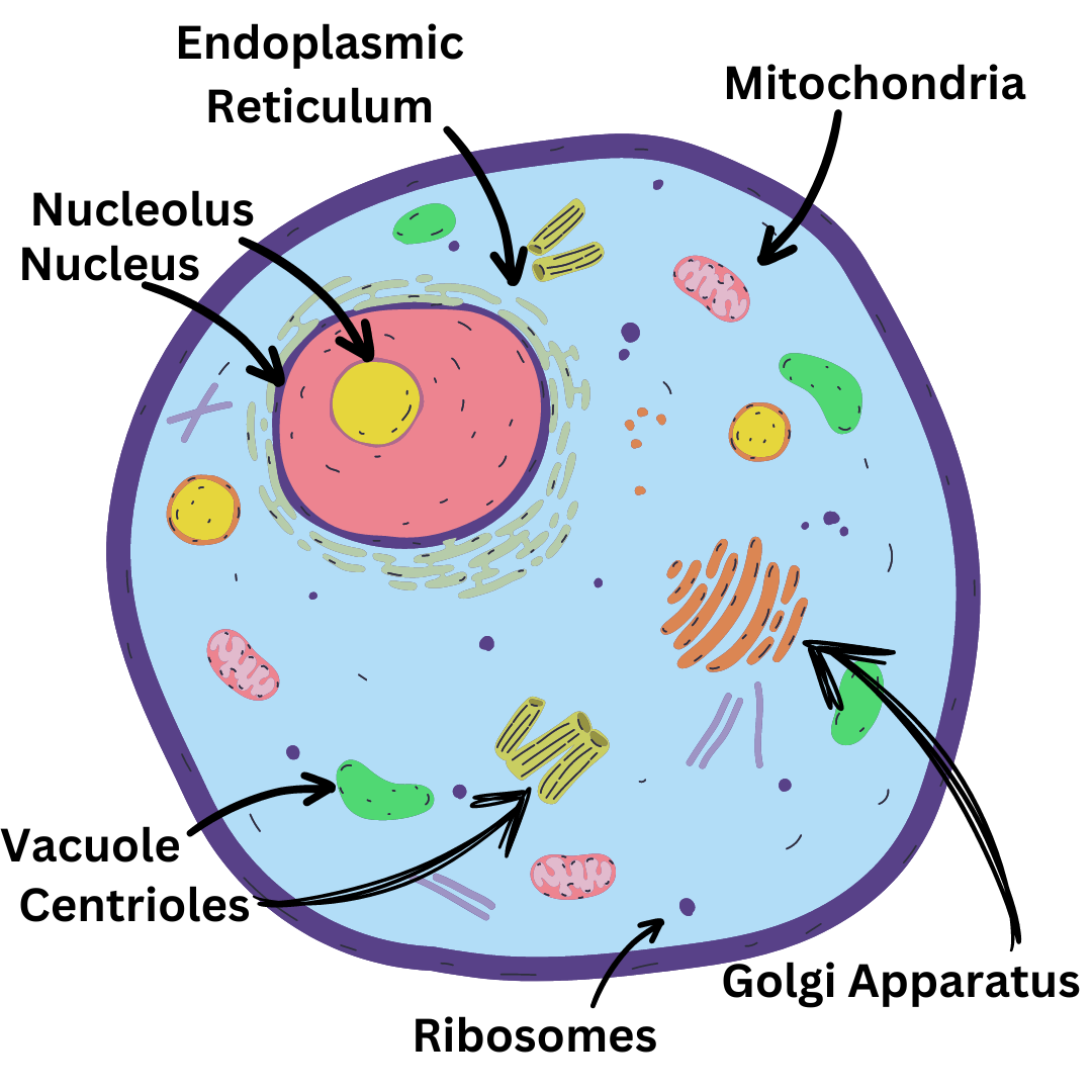
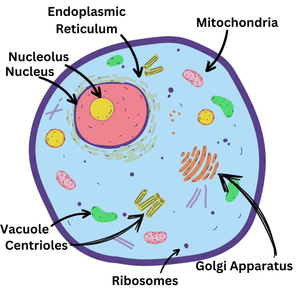
Animal cells are the basic units that make up all animals. Every animal, big or small, is made of these cells. They are tiny but essential. Inside each animal cell, there are many parts called organelles. Each organelle has a special job to do. Together, they help the cell stay alive and do its work.
Detailed Overview of Animal Cell Organelles
Let’s look at the different organelles found in animal cells and what they do. Below are the structure, location, and function of the organelles of an animal cell.
1. Nucleus
Structure:
The nucleus is large and round with a double membrane around it. Inside, it contains DNA and a smaller part called the nucleolus.
Location:
The nucleus is usually found in the center of the cell. It is one of the largest organelles in the cell.
Function:
It stores the cell’s DNA, which has the instructions for all the cell activities. The nucleolus inside the nucleus helps make ribosomes.
2. Mitochondira
Structure:
Mitochondria are small and bean-shaped. They have a double membrane: a smooth outer membrane and a folded inner membrane. The folds are called cristae.
Location:
Mitochondria are found throughout the cytoplasm of the cell. Few cells can have multiple mitochondria.
Function:
Mitochondria are the powerhouses of the cell. They produce energy by converting nutrients into ATP (adenosine triphosphate). This energy is used for various cell functions.
3. Ribosomes
Structure:
Ribosomes are small, and round but very important. They are made of proteins and RNA. They look like tiny dots under a microscope.
Location:
Ribosomes can be found floating in the cytoplasm. They can also be attached to the endoplasmic reticulum (ER), making it look rough.
Function:
Ribosomes make proteins. They follow instructions from the DNA to put together amino acids. Proteins help the cell grow and repair itself.
4. Endoplasmic Reticulum (ER)
Structure:
The endoplasmic reticulum (ER) is a large network of membranes. It comes in two types: rough ER and smooth ER.
A rough Endoplasmic Reticulum is a series of connected, flattened sacs that has ribosomes on its surface which makes it look rough. Smooth Endoplasmic Reticulum does not.
Location:
The ER is located around the nucleus. It is found in the cytoplasm.
Function:
The rough ER helps make and transport proteins. Ribosomes on the rough ER build proteins, which are then processed and sent to other parts of the cell or outside the cell.
The smooth ER makes and processes lipids (fats) and helps detoxify the cell.
5. Golgi Apparatus
Structure:
This organelle is a flattened-like, stacked pouches called cisternae. It resembles a stack of pancakes.
Location:
The Golgi apparatus is located in the cytoplasm, near the endoplasmic reticulum and the nucleus.
Function:
The Golgi apparatus is like a post office. It packages and ships proteins and lipids to where they are needed. It modifies them, sorts them, and then sends them to their final destinations, either inside or outside the cell. It ensures that all the cell’s materials are sent to the right places.
6. Lysosomes
Strucutre:
Lysosomes are small, round, membrane-bound organelles. They consist of digestive enzymes that help clean waste materials and cellular debris.
Location:
Lysosomes are found throughout the cytoplasm of the cell. They can vary in number depending on the cell type and its activity.
Function:
Lysosomes are like the cell’s cleanup crew. They digest dead or damaged organelles, food particles, and other unnecessary microorganisms. This keeps the cell clean and healthy.
7. Cell Membrane
Strucutre:
The cell membrane is a thin, flexible layer surrounding the cell. It is made of two layers of phospholipids with proteins embedded in them.
Location:
The cell membrane is found on the outermost boundary of the cell, separating the inside of the cell from the outside environment.
Function:
The cell membrane controls what enters and exits the cell. It acts as a barrier, protecting the cell and helping maintain its shape. It also allows nutrients to enter and water to leave.
8. Vacuoles
Strucutre:
Vacuoles are large, membrane-bound sacs within the cell. The vacuoles are filled with fluid and differ in size.
Location:
Vacuoles are found in the cytoplasm of the cell. Plant cells are usually large and central, while animal cells are smaller and scattered.
Function:
Vacuoles store nutrients, waste products, and other substances. They help maintain the cell’s shape and pressure by keeping the cell turgid. In plant cells, vacuoles also support growth and help with digestion.
9. Peroxisomes
Structure:
Peroxisomes are small, spherical organelles enclosed by a single membrane. They have a granular appearance due to the presence of enzymes.
Location:
Peroxisomes are scattered throughout the cytoplasm of the cell.
Function:
Peroxisomes help break down fatty acids and detoxify harmful substances. They produce hydrogen peroxide, which is then broken down by the enzyme catalase.
10. Centrioles
Structure:
Centrioles are cylindrical structures made of microtubules, typically found in pairs. They have a nine-triplet arrangement of microtubules.
Location:
Centrioles are located near the nucleus, within a region called the centrosome.
Function:
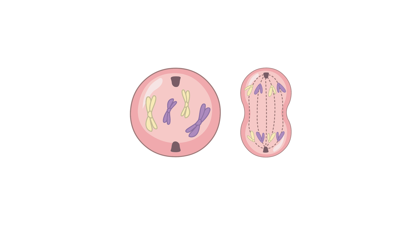
Centrioles play a key role in cell division by organizing microtubules. They help in forming the mitotic spindle during mitosis spindle during mitosis and meiosis.
11. Microtubule
Structure:
Microtubules are long, hollow tubes made of tubulin proteins. They are part of the cytoskeleton.
Location:
Microtubules extend throughout the cytoplasm and are anchored at the centrosome.
Function:
Microtubules provide structural support, help maintain cell shape, and facilitate intracellular transport.
12. Microfilaments
Stucture:
Microfilaments are thin, flexible threads made of actin proteins. They are part of the cytoskeleton.
Location:
Microfilaments are found throughout the cytoplasm, often forming a network just under the cell membrane
Function:
Microfilaments support cell shape, enable cell movement, and are involved in muscle contraction and cell division.
13. Cilia
Structure:
Cilia are short, hair-like projections on the surface of the cell. They are made of microtubules arranged in a 9+2 pattern.
Location:
Cilia are found on the exterior surface of some cells.
Function:
Cilia help with cell movement and the movement of fluids across the cell surface. They are important for functions like clearing mucus from the respiratory tract.
14. Flagella
Structure:
Flagella are long, whip-like projections from the cell surface, composed of microtubules arranged in a 9+2 pattern.
Location:
Flagella are found on the surface of some cells, usually in fewer numbers compared to cilia.
Function:
Flagella enable cell movement through a whip-like motion. They are crucial for the locomotion of the cells like sperm.
Difference Between Plant and Animal Cell
Animal and plant cells share many similarities but also have some key differences:
- Cell Wall: Plant cells have a cell wall made of cellulose that provides structure and protection. Animal cells do not have a cell wall; they only have a cell membrane.
- Chloroplasts: Plant cells can make their own food with the help of photosynthesis. Chloroplast in plants helps with this process. Whereas animal cells have no chloroplast.
- Shape: Plant cells are usually rectangular in shape due to the rigid cell wall. Animal cells are more flexible and can be round or irregular in shape.
- Vacuoles: Plant cells have large vacuoles in the middle of the cell which assist with water storage. Animal cells may have small vacuoles, but they are not as prominent as in plant cells.
- Centrioles: Animal cells have centrioles that play a role in cell division. Whereas, most of the plant cells lack centrioles.
FAQs about Animal Cell Structure
Here are some common questions about animal cell structure:
1. What is membrane-bonded organelle?
Membrane-bound organelles are cellular structures surrounded by their own lipid membranes. These organelles are enclosed by one or more membranes, separating their internal environment from the rest of the cell. This membrane-bound structure allows them to perform specialized functions within the cell, such as processing proteins, generating energy, or storing substances, in a controlled and isolated environment. For example, nuclei, mitochondria, etc.
2. How does the Golgi Apparatus help the cell?
The Golgi Apparatus packages and ships proteins and lipids to where they are needed in the cell
3. Why are Lysosomes important for the cell?
Lysosomes are important because they digest waste materials and recycle old cell parts, keeping the cell clean and healthy.
Conclusion
Understanding animal cell structure is fundamental to grasping how cells operate and sustain life. Each organelle within the cell – such as the nucleus, mitochondria, ribosomes, endoplasmic reticulum, Golgi apparatus, lysosomes, and others – plays a crucial role in maintaining cellular functions and health.